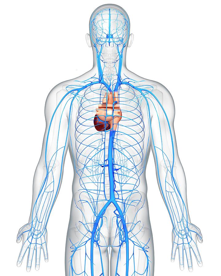
Dr Kim Wilson Lab 6 Circulatory System Part 4 Veins Biology Diagrams At any time, nearly 70% of the total volume of blood in the human body is in the veins. [3] The anatomy of the veins of the heart is very variable, but generally it is formed by the following veins: heart veins that go into the coronary sinus: the great cardiac vein, Trunk Veins of the trunk converge from the thorax, abdomen, and pelvis towards the heart. Deoxygenated blood from the thorax ultimately drains into the superior vena cava (SVC).The major thoracic tributaries of the SVC include the: azygos venous system, pulmonary veins, internal thoracic vein and cardiac veins. Venous blood from the abdomen and pelvis is drained by the inferior vena cava.

Structure of Veins. Veins are thin-walled valves containing blood vessels. Anatomically, veins have the same three layered walls as arteries that are the tunica externa, the tunica media, and the tunica intima; however, they have considerably lesser amounts of smooth muscles making them thinner than the walls of the arteries. This thinner wall makes the veins more flexible and allows the veins

What Are Veins and How Do They Work? Biology Diagrams
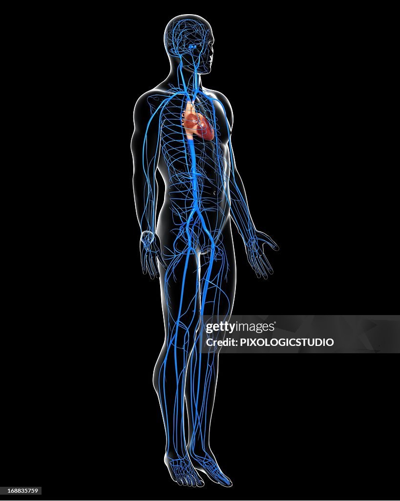
Human Vascular System. Veins (blue) and Arteries (red). SEBASTIAN KAULITZK/Science Photo Library/Getty Images. Veins can be categorized into four main types: pulmonary, systemic, superficial, and deep veins. Pulmonary veins carry oxygenated blood from the lungs to the left atrium of the heart.; Systemic veins return oxygen-depleted blood from the rest of the body to the right atrium of the heart.
We'll explain the basic structure of a vein before diving into different types of veins and their functions. Explore the venous system with an interactive diagram and learn some tips for

Veins: Definition, Structure, Types, Functions, Diseases Biology Diagrams
Internal thoracic veins: Superior epigastric veins, Musculophrenic veins, and Anterior intercostal veins are the internal thoracic veins.Supreme intercostal vein Azygos Vein: The azygos vein is a major vein on the right side of the body that drains blood from the posterior thoracic wall and empties into the Superior vena cava.