Prophase chromosomes are free to exchange neighbors a Simulation the Biology Diagrams It occurs in several stages, each of which consists of a stereotyped set of changes in cell contents and structure. In this article, we will look at the stages of mitosis and its clinical relevance. To help with this, at the start of prophase, chromatin begins condensing into chromosomes. In addition, mitotic spindles begin to form. In prophase, the chromatin condenses into discrete chromosomes. The nuclear envelope breaks down and spindles form at opposite poles of the cell. Prophase (versus interphase) is the first true step of the mitotic process. During prophase, several important changes occur: Chromatin fibers become coiled into chromosomes, with each chromosome

During prophase, the replicated chromosomes (each consisting of two sister chromatids) condense and are recognizable under the microscope. Survivin is responsible for targeting the CPC to chromatin by associating with histone modifications, Topoisomerase IIα induces a conformational change that places the G-segment within its active
TeachMePhysiology Biology Diagrams
phase chromatin changes occur at nuclear pores, on the inner surfaceofthenuclearenvelope, andmoststrikinglyinthenucle-olus. There, proteins involved in rRNA processing move away their proximity to chromatin during prophase. However, about 20% remain relatively unaffected (Figures 3D and 3E; orange cluster). This behavior was well reproduced During prophase, the chromatin condenses into visible chromosomes. Each chromosome consists of two sister chromatids. These are connected at a region called the centromere. The mitotic spindle also begins to form during this stage. Understanding the carbon cycle is essential for addressing climate change and sustaining ecosystems. Through

Similar changes were seen in wild-type cells in a previous study. 4 Then, during prophase (−20 to 0 min relative to NEBD) and prometaphase (after NEBD), more cells showed the resolved configuration (4 dots; 2 of each color; pink pattern) and the folded configuration (2 pairs of colocalized red and green dots; red pattern) in both cell lines
+Centrioles+move+to+poles..jpg)
Mapping the invisible chromatin transactions of prophase chromosome ... Biology Diagrams
The process is longer due to the phases of prophase which takes place in two phases i.e prophase I and prophase II. Prophase I is quite complex which involves the pairing up of the homologous chromosomes and the exchange of genetic information. It defines the difference between mitosis and meiosis. Prophase II is very similar to the mitotic
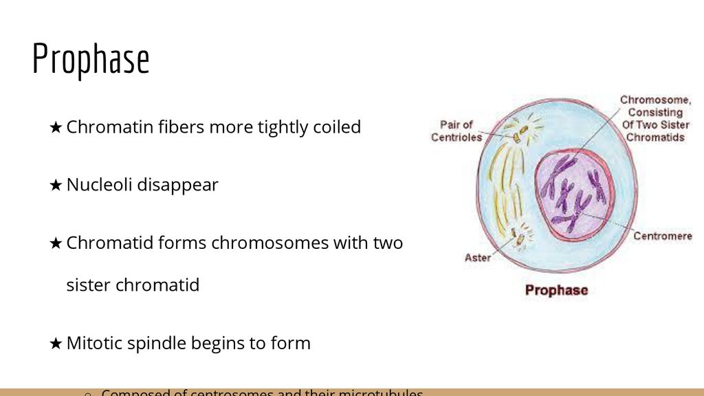
Explore the essential stages of prophase in mitosis, focusing on chromosome condensation, spindle formation, and nuclear envelope breakdown. Specific chemical changes to histones, such as phosphorylation, play a role in altering the chromatin structure, making it more compact. These modifications are tightly regulated by various enzymes
